The Magic of Tensegrity
Imagine a world where strength doesn’t rely on rigid walls but on a delicate balance between tension and compression. This is the magic of tensegrity—a principle that shapes everything from futuristic architecture to the very cells in our bodies. But it doesn’t stop there.
How Tensegrity and Mechanotransduction work in harmony
Mechanotransduction, the process by which cells sense and respond to mechanical forces, opens new possibilities when applied to revolutionary materials like SP1KE™. Just as your body adjusts to pressure and movement, SP1KE™ utilizes tensegrity—stability achieved through continuous tension and compression in a pre-stressed network (Ingber, 2008b). By maintaining a pre-stressed state, SP1KE™ effectively balances and distributes various loads, ensuring stability and setting it apart from other products.
SP1KE™ technology harnesses the principles of tensegrity and mechanotransduction
Enter SP1KE™ technology, developed with insights from Dr. N. Apostolopoulos, Ph.D. This cutting-edge innovation basically harnesses the principles of tensegrity and mechanotransduction, to create products that not only offer comfort and protection, but also interact with your body in ways you never thought possible.
SP1KE™ technology adapts to tension and compression
SP1KE™ by Vigurus Technologies revolutionizes human interaction with the environment (sitting, standing, lying) by adapting to various forces like tension, compression, shear, bending, and torsion. Furthermore, its patented design redistributes loads three-dimensionally, enhancing the connection between the body and structure while maintaining integrity.
SP1KE™ mimics the interaction of cells within tissues, with its geometric nodes functioning similarly to the extracellular matrix (ECM) in the body.
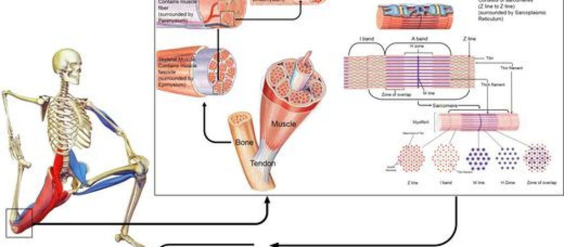
Just as cells rely on ECM and integrins for growth and function, SP1KE™ adapts to applied forces, triggering a biochemical response through mechanotransduction. This dynamic adaptability reflects the way muscles and ECM respond to physical stress, above all, showcasing SP1KE™’s explicit ability to adjust to various loads.
N. Apostolopoulos, Ph.D. and founder of MicroStretching® says Sp1ke heightens the mechanotransduction response
As the founder and developer of Stretch Therapy & the technique of microStretching®, I have always been interested in the response of stretching intensity and its effect on inflammation/inflammatory response. As I have shown, I believe that the use of Sp1ke, in conjunction with microStretching only heightens the mechanotransduction response. Accordingly, Sp1ke has huge applicability for the recovery of muscle & to provide a therapeutic aide to individuals suffering from muscle injury, and pathological conditions associated with inflammation (i.e., osteo-arthritis).
Curious about how this breakthrough could redefine seating, sports gear, and even therapeutic devices? Dive into the fascinating world where science meets invention, and discover the truth about how SP1KE™ is changing the game. Check out www.vigurus.com and particularly Sp1ke insoles that offer an undeniable and marked difference in postural alignment, balance and muscle recovery.
REFERENCES
GANS, C. & ABBOT, S. G. 1991. Muscle architecture in relation to function. J Biomech, 24, 53-65.
HUMPHRIES, M. J. 2000. Integrin structure. Biochem Soc Trans, 28, 311-339.
HYNES, R. O. 2002. Integrins: bidirectional, allosteric signaling machines. Cell, 110, 673-687.
INGBER, D. E. 1998. The architecture of life. Sci Amer, 278, 48-57.
INGBER, D. E. 2006. Cellular mechanotransduction: putting all the pieces together again. FASEB, 20, 811-827.
INGBER, D. E. 2008a. Tensegrity-based mechanosensing form macro to micro. Prog Biophys Mol Biol, 97, 163-179.
INGBER, D. E. 2008b. Tensegrity and mechanotransduction. J Bodyw Movem Ther, 12, 198-200.
KOMULAINEN, J., TAKALA, T. E. S., KUIPERS, H. & HESSELINK, M. K. C. 1998. The disruption of the myofibre structures in rat skeletal muscle after forced lengthening contractions. Pflugers Arch – Eur J Physiol, 117, 29-35.
MARTINS, W. R., CARVALHO, M. M., MOTA, M. R., CIPRIANO, G. F. B., MENDES, F. A. S., DINIZ, L. R., JUNIOR, G. C., CARREGARO, R. L. & DURIGAN, J. L. Q. 2013. Dicutaneous fibrolysis versus passive stretching after articular immobilisation: muscle recovery and extracellualr matrix reodelling. OA Med Hypo, 1, 17-22.
MEREDITH JR, J. E. & SCHWARTZ, M. A. 1997. Integrins, adhesion and apoptosis. Trends Cell Biol, 7, 146-150.
RALPHS, J. R., WAGGET, A. D. & BENJAMIN, M. 2002. Actin stress fibres and cell-cell adhesion molecules in tendons: organisation in vivo and response to mechanical loading of tendon cells in vitro. Matrix Biol, 21, 67-74.
RUOSLATHI, E. & REED, J. C. 1994. Anchorage dependence, integrins, and apoptosis. Cell, 77, 477-478.


